Material: 1 bistoury; 2 kidneys of mammal; 1 dissecting tray; 1 tweezer; 1 dissecting needle; gloves (latex); squirt of distilled water ; cleaning paper; pins.
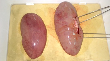
In the image above, we can see the two kidneys before they were dissected. It is possible to identify the capsule, a smooth, fibrous and transparent membrane, that involves the kidney and the hilum.
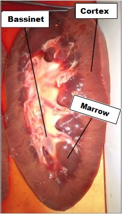
In this second image, it's possible to identify the 3 main parts of the inside of the kidney, after being dissected. It is possible to identify the medullary zone, which contains the renal pyramids; the bassinet, an upper portion, next to the ureter, which contains the urine; and the cortex zone, a peripheral layer, which goes from the capsule until the renal pyramids.
